The "Segmentation and Intensities Analysis" tool processes several T1 images for different sessions of the same subject to extract T1 values for regions of interest, such as dentate or globus pallidus, and to normalize by CSF or brainstem T1 most common value.
1. Show Results
Open My Analysis to see analysis results you have already run.
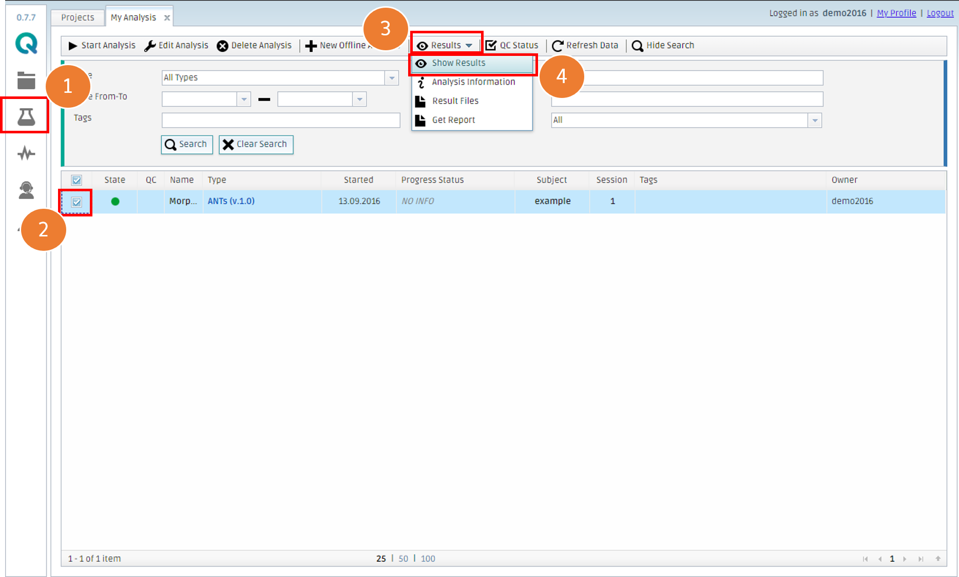
- Click My Analysis
- Check an analysis you want to browse (Pick a Green circle, which means a completed analysis, in the second column)
- Click Results
- Click Show Results
Alternatively, you can double-click the row of the analysis.
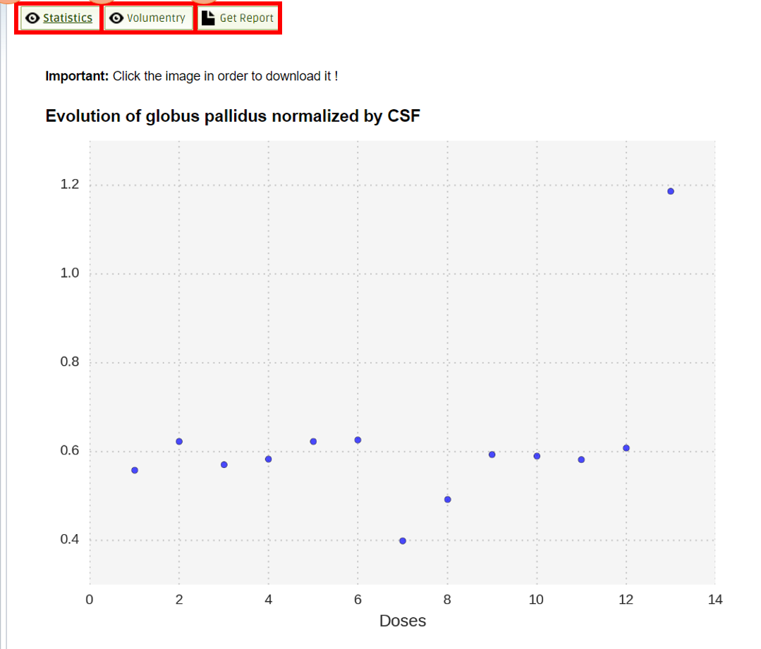
- See statistics of the cohort analysis
- See the result of volumetry analysis
- Download a formatted document in PDF
2. Download output files
You can download specific values on regions of interest.
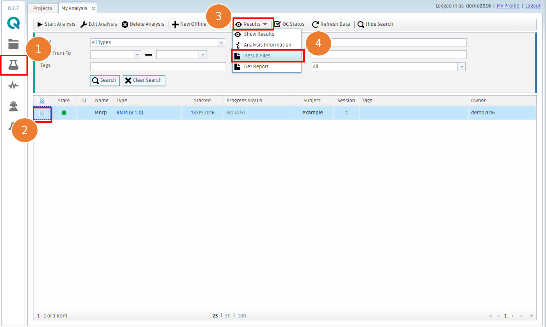
- Go to My Analysis
- Check an analysis you want to browse (Pick a Green circle, which means a completed analysis, in the second column)
- Click Results
- Click Result Files
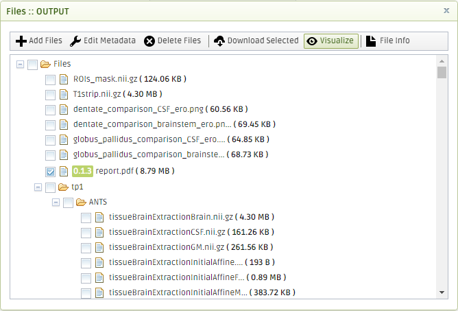
You can select one or more files and click Download Selected to get the files. The output files have the following name structure.
- T1_ {region_name}.nii.gz : These files are T1 extracted regions. The only voxels that are not zero are the ones in the region of interest, values from T1 contrast.
- ROIs_mask.nii.gz: The values for these regions are NOT from any atlas. This image is used only for the visualization of the results. The values are extracted from a color lookup table that has associated those values to the colors shown in the Show Results panel. Binary mask with 5 regions:
- brainstem
- globus pallidus left
- globus pallidus right
- dentate left
- dentate right
- T1_BS_CEREBELLUM.nii.gz: Brainstem and cerebellum segmented from T1.
- T1_acpc.nii.gz: Preprocessed T1 image aligned to acpc
- T1strip.nii.gz: Skull-stripped T1 image.
- tissueSegmentation.nii.gz: Final tissue segmentation image.
- T1mask.nii.gz: Binary mask image of brain non-CSF tissues
- atlas_registered.nii.gz: Atlas labels wrapped to subject T1.
- full_labeled.nii.gz: Complete parcellated labels image.
- labeled.nii.gz: Filtered labels image used for volumetry.
- ANTS: Folder with full output of the ANTs tool
The other timepoints have the registered masks. The tool takes the last timepoint T1strip and registers it to each time point and then the registration is applied for all the T1_{region_name}.nii.gz file.
Create free account now!
