Segments T2 hyperintense lesions in FLAIR images, such as those found in multiple sclerosis (MS). Although originally developed for MS, it has also proven to be useful to segment lesions caused by other diseases, such as Alzheimer's (AD).
It makes use of the ST tool , an open source toolbox developed by a group of institutions, including the Technical University Munich, Ludwig Maxmilians University and Friedrich Schiller University.
This implementation uses the lesion prediction algorithm (LPA) which does not require any user-defined parameter Schmidt 2017 . This fast and sensitive algorithm was trained by a logistic regression model with the data of 53 MS patients with severe lesion patterns.
You can use LST tool for lesion segmentation of MS, AD, and other diseases. You may also use the outputs to evaluate your own lesion segmentation algorithm by comparing your results with the results of the LST tool.
The tool also calculates the lesion volume and the lesion count.
Lesion Segmentation Tool output examples
FLAIR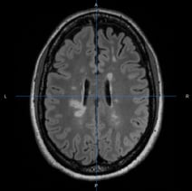 |
Lesion probability map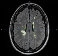 |
Lesion segmentation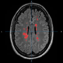 |
Required inputs:
- T2-FLAIR: anatomical 3D image with hyper-intense lesions
Isotropic resolution recommended
Must be labelled as 'T2' modality
Minimum input requirements:
- For optimal result reliability, isotropic resolution is recommended
- Recommended resolution: 1 mm isotropic.
- Minimum reliable resolution: 2mm isotropic.
Settings:
- Reorient to STD
-Reorient to STD (checkbox) If checked reorient image to standard (Default checked) - Binarize probability maps
-Binarize threshold (float) (Default 0.0)
Outputs:
- Images:
- T2.nii.gz: the input T2 image used in the tool.
- lesions_label.nii.gz: lesions labeled with unique identifiers.
- ples_lpa_mT2.nii.gz: probability map of lesion voxels.
- ples_lpa_mT2_bin.nii.gz: output segmented lesion mask.
- Datasheets:
- detail_lesions_info.csv: Volume of each lesion
- global_lesions_info.csv: Total lesion volume
- Other:
- hist_volumes_lesions.png: Histogram lesion volumes image
- report.pdf: PDF report (tags: report)
Typical execution time:
- 10 minutes.
References:
- Lesion prediction algorithm: Schmidt 2017
Create free account now!
