Generates a whole-brain structural connectome, characterizing the white matter connections between grey matter areas. Combining the capabilities of T1-based morphology and dMRI-based tractography, the structural connectome tool enables the quantification of brain connectivity between different the cortical and sub-cortical region of interest (ROIs).
|
T1-based ROI Parcellation |
|
dMRI-based Tractography |
Connectivity matrix circular plot |

The full workflow includes:
-
DICOM files to NIfTI conversion and reorients to standard. The workflow file filter sends the dicom or nifti files required for the workflow to run and the box converts the dicom to niftis, extracts the necessary files (if possible) to do the dmri preprocessing step (eddy) and gradient tables. If nifti files or gradient tables are found in the input, it just does the reorient to standard step and outputs the files.
-
dMRI preprocessing includes denoising, gibbs ringing correction, eddy correction, motion correction, bias field correction, distortion correction (if gradient field maps are available) and ACPC alignment.
- Gets the brain mask and computes the tensor from the diffusion image, plus derives different scalar measures: FA, AD, MD, RD, Cl, Cs, Cp.
-
Runs ants cortical volumetry script over T1 image to extract the tissue segmentation file.
-
Applies an atlas to the segmented mask of the cortex (gray matter + subcortical gray matter + brainstem + cerebellum)
-
Align the diffusion image to the T1
-
Estimate the diffusion model. Does nothing if the data is DTI. Runs constrained spherical deconvolution model, multi-tissue and multi-shell processing is available.
-
Perform probabilistic or deterministic tractography over the diffusion model, ACT (using the 5tt format from mrtrix) is available.
-
Estimates the connectome using the tractography and the registered labels. Outputs the normalized connectomes by volume and/or fiber length. Connectome analysis via different graph-based measures: characteristic path length, density, node degree, assortativity, modularity, efficiency, and clustering coefficient (local and global).
Required inputs:
- T1: anatomical 3D image
Isotropic resolution recommended
Must be labeled as 'T1' modality - dMRI: diffusion 4D image (DTI or HARDI).
Isotropic resolution recommended.
Must be labeled as 'DTI' or 'HARDI' modality. - Gradients: gradient table (optional if dMRI is DICOM).
MRtrix format supported: file must have '.b' extension and be labeled as 'gradient_table'.
FSL format supported: files must have '.bvec' and '.bval' extensions and be labeled 'bvec' and 'bval'.
Minimum input requirements:
- Image must contain full brain DWI dataset.
- For optimal result reliability, an isotropic resolution is highly recommended
- T1 recommended resolution: 1 mm isotropic.
- T1 minimum reliable resolution: 2mm isotropic.
- dMRI recommended resolution: 2 mm isotropic.
- dMRI minimum reliable resolution: 3 mm isotropic.
- T1 recommended resolution: 1 mm isotropic.
- If DTI image must have at least 7 volumes (1 b0 and 6 diffusion volumes).
- If HARDI image must have at least 21 volumes (1 b0 and 20 diffusion volumes).
- For optimal preprocessing reliability, 1 b0 image is recommended every 10 gradient volumes.
Optional inputs:
- GFMs: Gradient field-maps, recommended (magnitude image and phase image).
Same resolution and FOV as dMRI image recommended.
Files must be labeled 'gfm_magnitude' and 'gfm_phase' respectively.
Files must additionally be labeled 'dti' or 'hardi' depending on dMRI modality label.
Settings
ACPC_t1:
- Check when data is skull stripped (checkbox)
Enable this if the input data is skull stripped (default unchecked) - Change image resolution (checkbox)
Changes the voxel size to isotropic resolution, defined in the next setting (default checked) - Isotropic voxel final size (mm) (integer)
Define new isotropic resolution for the T1 image (default: 1)
DCM2NII:
- Preferred DICOM to NIfTI conversion tool (drop-down selection)
The selected tool will be tried first to convert DICOM to NIfTI. If the conversion fails, the other options will be tried sequentially until a successful conversion.- DCM2niix (Default)
- Mrtrix
- DCM2nii
- diffunpack
ANTS
- Compute thickness map (checkbox)
Perform additional cortical thickness on T1 (default unchecked)
PREPROCESSING
- Do denoising (checkbox)
(default checked) - Do Gibb ringing correction (checkbox)
(default checked) - Do Eddy (checkbox)
(default checked) - Do Fugue (checkbox)
(default checked) - Use manual diffusion acquisition parameters (checkbox)
If checked uses the parameters input manually in the following settings (Default unchecked). - Echo Spacing (s) (float)
The time between echoes in an echo planar image (EPI) (Default 0.0003ms) - Echo Factor (integer)
Also called Echo Train Length (ETL), the number of echoes acquired in a single repetition time (TR) (default 100) - Acceleration Factor (integer)
The number of EPI lines acquired in parallel (Default 1) - Echo Difference (s) (float)
(only needed if GFMs are provided), the difference in Echo Time (ET) between GFM acquisitions. (default 0.00246 for SIEMENS acquisitions) - AC-PC alignment (checkbox)
(default unchecked) - Resample dMRI image (checkbox)
(default checked) - dMRI isotropic resampling resolution (mm) (float)
Resamples the dMRI image to the indicated isotropic resolution in mm. (Default 2 mm)
MASK_ALIGNMENT
- Compute non-linear registration EPI to T1 (FA input required) (checkbox)
(default checked)
ATLAS
- Brain atlas for parcellation (drop-down selection)
The selected tool will be tried first to convert DICOM to NIfTI. If the conversion fails, the other options will be tried sequentially until a successful conversion.- DKT40 (Mindboggle-101) (default)
- AAL
MODEL
- Tracking algorithm type (drop-down selection)
- Constrained Spherical Deconvolution (CSD) (default)
- Tensor
- Multi-tissue constrained spherical deconvolution
TRACTO
- Max. spherical harmonic order (drop-down menu):
- 4
- 6 (default)
- 8
- 10
- Tracking algorithm type
- DET deterministic
- PROB probabilistic
- Number of streamlines (integer)
Number of streamlines to generate (default 1M) - Step size (mm) (0 for adaptative default) (float)
(default 0) - Use Anatomical constrained tractography (checkbox)
Only works when Morphology results for ACT are passed as input (default checked) -
Stopping angle (º) [0 for adaptive default] (integer)
(default 0) - Min. streamline length (mm) [0 for adaptive default] (integer)
(default 0) - Max. streamline length (mm) [0 for adaptive default] (integer)
(default 0) - Min. FA/FOD cutoff (float)
(default 0)
Output files
We explain the output files for each box. The modalities are shown in colored boxes and the tags are shown with outlined boxes. Each element represents a relevant file from the container, more might be present but they are not as important.
ACPC_t1
- T1HEAD T1 image aligned to ACPC
- REPORTIMAGE PNG image for QC
DCM2NII
- T1 : T1 NIfTI file
- HARDI / DTI : dMRI NIfTI file
- HARDI / DTI:
- HARDI / DTI ACQ_PARAMS: acquisition parameter file for eddy preprocessing automatically extracted from dicom file
- HARDI / DTI BVAL: BVAL file FSL format
- HARDI / DTI BVEC: BVEC file in FSL format
- HARDI / DTI GRADIENT_TABLE: gradient table in mrtrix format
- HARDI / DTI INDEX: index file for eddy preprocessing automatically extracted from dicom file
ANTS
- T1 HEAD Full T1 head image
- T1 BRAIN Skull stripped T1
- T1 MASK Binary skull stripped brain mask
- TISSUE_SEGMENTATION 6-tissue segmentation image (1: CSF, 2: gray matter, 3: white matter, 4: deep gray matter, 5: brainstem, 6: cerebellum)
- CSV Volumetry information for ICV and each segmented tissue.
PREPROCESSING
- HARDI / DTI : dMRI NIfTI file processed with the steps selected in the settings
- GRADIENT_TABLE: gradient table in mrtrix format
- AVERAGE_B0S: Average b0s from the processed dMRI image
- REPORT: PDF report of the box
MEASURES
- TENSOR: Diffusion tensor data
- AVERAGE_B0S: Average b0s from the processed dMRI image
- MASK: dMRI binary brain mask
FA
- SCALAR AD: AD scalar file
- SCALAR CL: Cl scalar file
- SCALAR CP: Cp scalar file
- SCALAR CS: Cs scalar file
- SCALAR FA: FA scalar file
- SCALAR MD: MD scalar file
- SCALAR RD: RD scalar file
- REPORT : PDF report of the box
ATLAS
- ATLAS_INFO : Text file with atlas information
- LABELS : Labeled cortical brain
- REPORT : PDF report of the box
- Vectorized data folder
- VECTOR : Volumetry information in a vector format
MASK_ALIGNMENT
- HARDI / DTI : dMRI NIfTI file aligned to the anatomical image
- SCALAR AD: AD scalar file aligned to the anatomical image
- SCALAR CL: Cl scalar file aligned to the anatomical image
- SCALAR CP: Cp scalar file aligned to the anatomical image
- SCALAR CS: Cs scalar file aligned to the anatomical image
- SCALAR FA: FA scalar file aligned to the anatomical image
- SCALAR MD: MD scalar file aligned to the anatomical image
- SCALAR RD: RD scalar file aligned to the anatomical image
- ACT : ACT volume (5tt format) for connectome
- AVERAGE_B0S: Average b0s from the processed dMRI image aligned to the anatomical image
- GRADIENT_TABLE: gradient table in mrtrix format
- LABELS : Labeled cortical brain for connectome
- MASK: dMRI binary brain mask aligned to the anatomical image
- TISSUE_SEGMENTATION 6-tissue segmentation image for connectome
MODEL
- REPORT PDF report of the box
HARDI case
- case CSD
-
CSD CSD model
- RESPONSE Response file
- MASK Mask used to create the response file
-
- case msmt
- MSMT MSMT white matter model
- FODCSF MSMT CSF model
- FODGM MSMT GM model
- RESPONSECSF Response file of the CSF
- RESPONSEGM Response file of the GM
- RESPONSEWM Response file of the WM
- MASKCSF Mask used to create the response file of the CSF
- MASKGM Mask used to create the response file of the GM
- MASKWM Mask used to create the response file of the WM
DTI case
- DTI Input file
TRACTO
- TRACKS: streamlines computed from diffusion data. When using probablilistic algorithm it will be also tagged with PROB , but if deterministic algorithm is used, it will be tagged with DET
- TRACKSDOWNSAMPLED : streamlines computed from diffusion data downsampled for visualization purposes
CONNECT
- ASSIGNMENTS_FINAL : File for connectivity per-track assignment in MRtrix format, each row contains the id of the source and the target anatomical label per each fiber.
- ATLAS_INFO : Text file with atlas information of the connectivity regions
- TRACKS : Connected tracks from original tractography
- TRACKSDOWNSAMPLED : Tractography for visualization
- REPORT : PDF report of the box
- LABELS : Labeled cortical brain for connectome
- CONNECTOME : Connectome matrix
- CONNECTOMENORM_VOLUME: Connectome matrix normalized by volume
- CONNECTOMENORM_LENGTH: Connectome matrix normalized by length
- CONNECTOMENORM_VOLUMENORM_LENGTH: Connectome matrix normalized by volume and length
- vectorized_data folder
- VECTOR : Vectorized connectome matrix
- VECTORNORM_VOLUME: Vectorized connectome matrix normalized by volume
- VECTORNORM_LENGTH: Vectorized connectome matrix normalized by length
- VECTORNORM_VOLUMENORM_LENGTH: Vectorized connectome matrix normalized by volume and length
Typical execution time:
- 2 hours.
Additional details:
- The connectome files are plain CSV text files. It has the shape of a NxN matrix, being N the total number of regions of interest. Each element of the file is a “cell” and it represents a connection. Each row and column of the file represents a region of interest: the n-th row and the n-th column represent the same region of interest. We are only interested in the connectivity between different regions, and that is why the diagonal of the matrix is all zero.
- The regions of interest analysed in the connectome are the same as the ones found in the volumetric.csv file and they are in the same order in the matrix as in the volumetric file. The regions of interest start after the BPF.
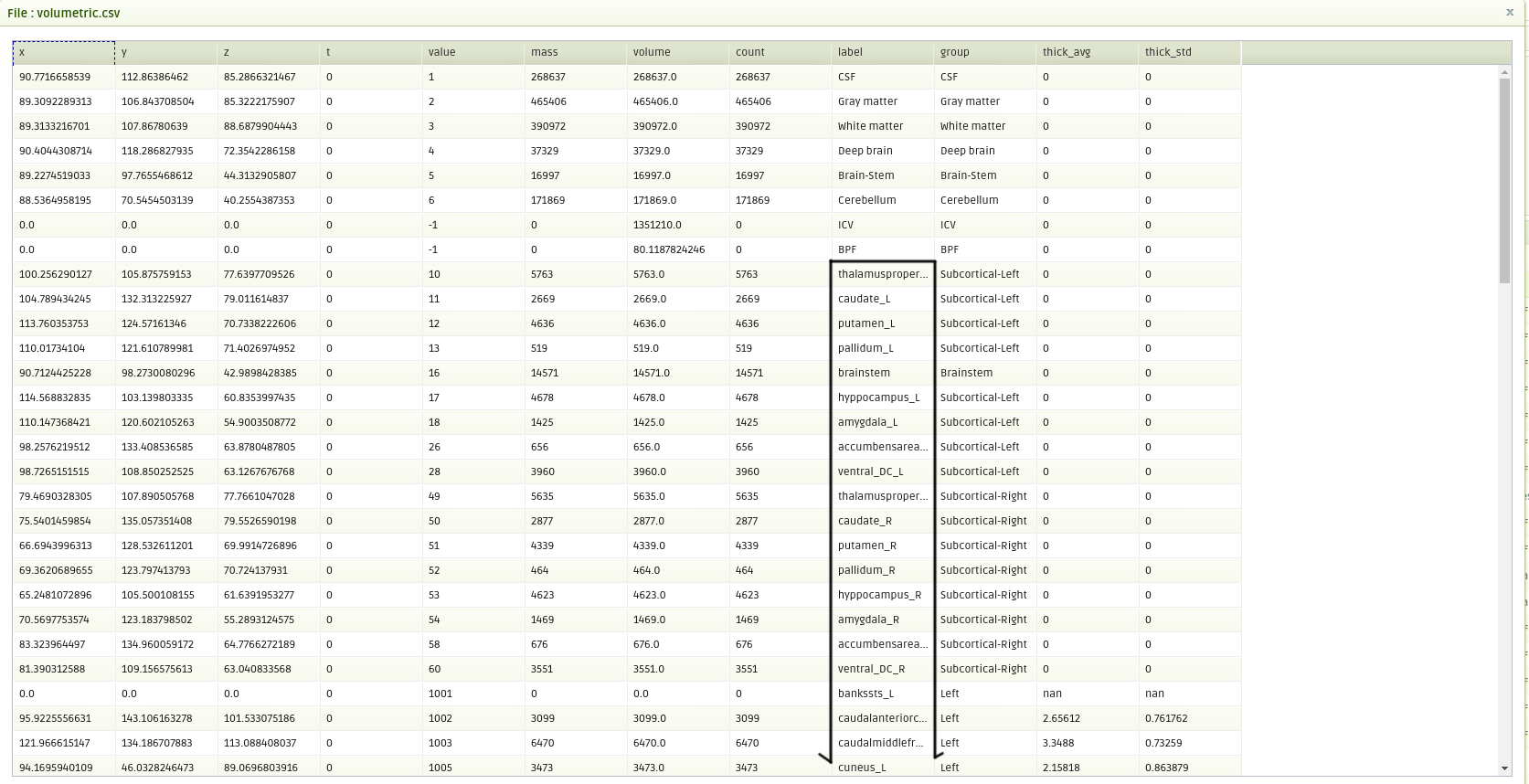
Region names found in volumetric.csv from BPF
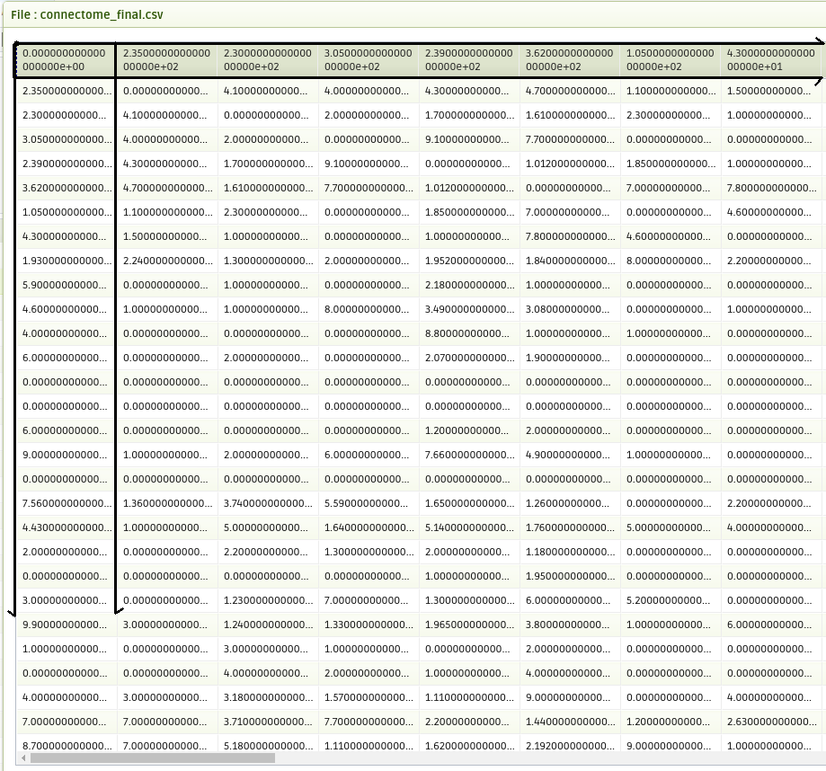
Connectivity follows the same order as in volumetric.csv
Create free account now!

References
dMRI PREPROCESSING
-
DWI gradient table check [Ben Jeurissen et al. 2014]
- Subject motion & Eddy correction: [Andersson and Sotiropoulos 2016]
- Distortion correction: [Jezzard and Balaban 1995].
ACPC alignment:
-
ANTs registration: [Avants et al. 2008]
- MNI152 Atlas: [Grabner et al. 2006]
-
FLIRT registration: [Jenkinson et al. 2002]
dMRI tensor and scalar measures
- MRtrix tools: [Tournier et al. 2012]
- Tensor estimation: [Veraart et al. 2013]
- Scalar measures: [Basser et al. 1994], [Westin et al. 1997] ("Geometrical diffusion measures for MRI from tensor basis analysis". Proc Intl Soc Mag Reson Med).
T1 segmentation and cortical labels
-
ANTs script: [Tustison et al. 2014]
-
ANTs registration: [Avants et al. 2008]
-
Atropos tissue segmentation: [Avants et al. 2011]
-
DiRECT cortical thickness: [Das et al. 2009]
-
OASIS-30 tissue priors: [Landman and Warfield 2012] ("MICCAI 2012 workshop on multi-atlas labeling." Medical image computing and computer-assisted intervention conference)
-
DKT40 Mindboggle 101 Atlas: [Klein and Tourville, 2012]
-
AAL Atlas: [Tzourio-Mazoyer et al. 2002], [Rolls et al. 2015]
-
MNI152 Atlas: [Grabner et al. 2006]
MASK_ALIGNMENT
- ANTs registration: [Avants et al. 2008]
Diffusion MODEL
- msmt_5tt [Ben Jeurisen et al. 2014]
- CSD [J-Donald Tournier et al. 2007]
- fod2dec: Two posters [T. Dhollander et al., 2015] [T. Dhollander et al. 2015]
TRACTO
- iFOD1 or SD_STREAM: [J-Donald Tournier et al. 2012]
- iFOD2 poster [J-Donald Tournier et al. 2010]
- Tensor_Det [Basser, P.J. et al. 2000]
- Tensor_Prob [Jones, D. et al. 2008]
- act, -backtrack, -seed_gmwmi [Robert E. Smith et al. 2012]
CONNECT
- Connectome normalization [Hagmann P. et al. 2008]