This workflow segments the main structures of gliomas from MR images and includes a manual correction step to further refine the segmentation labels.
 |
 |
 |
 |
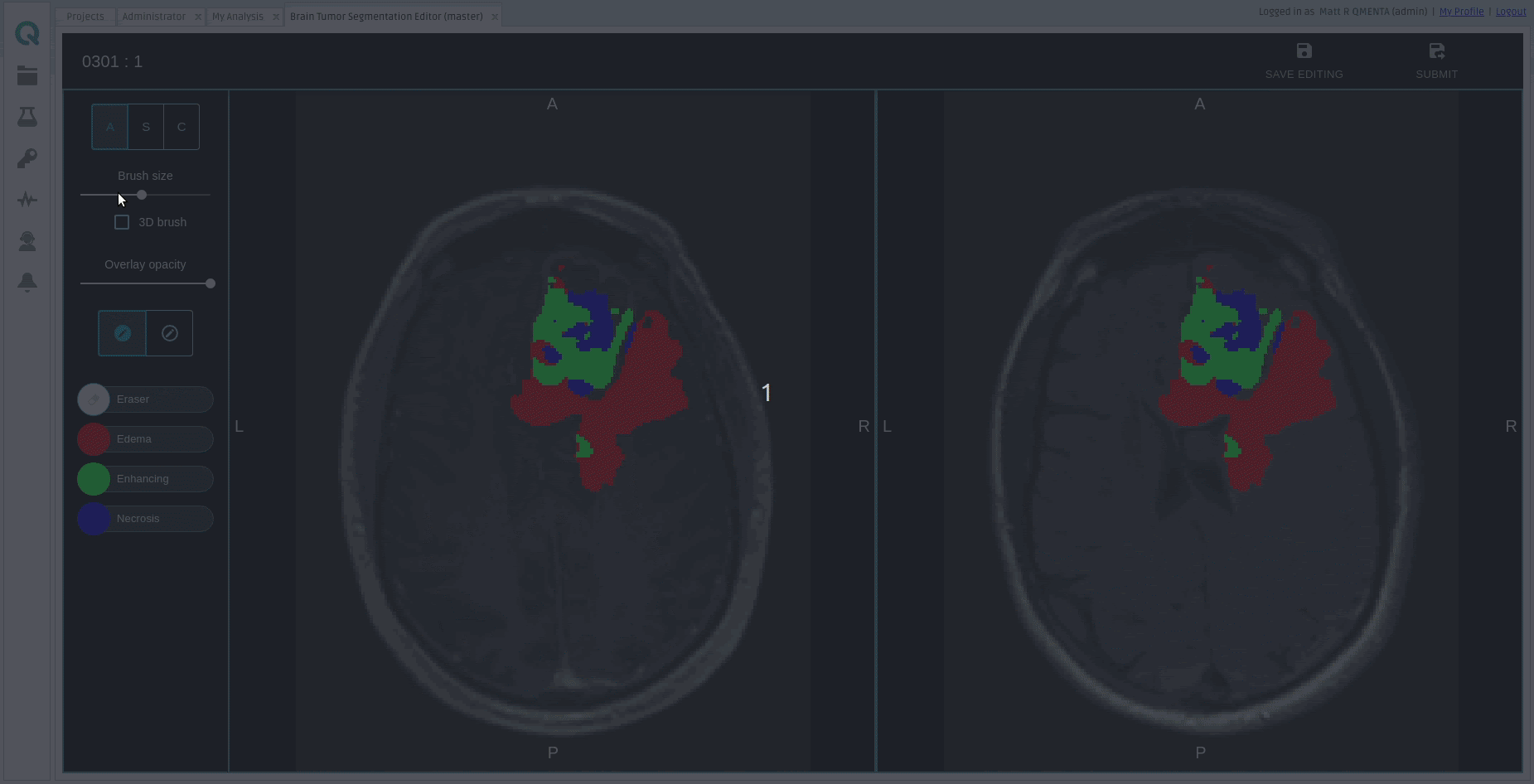
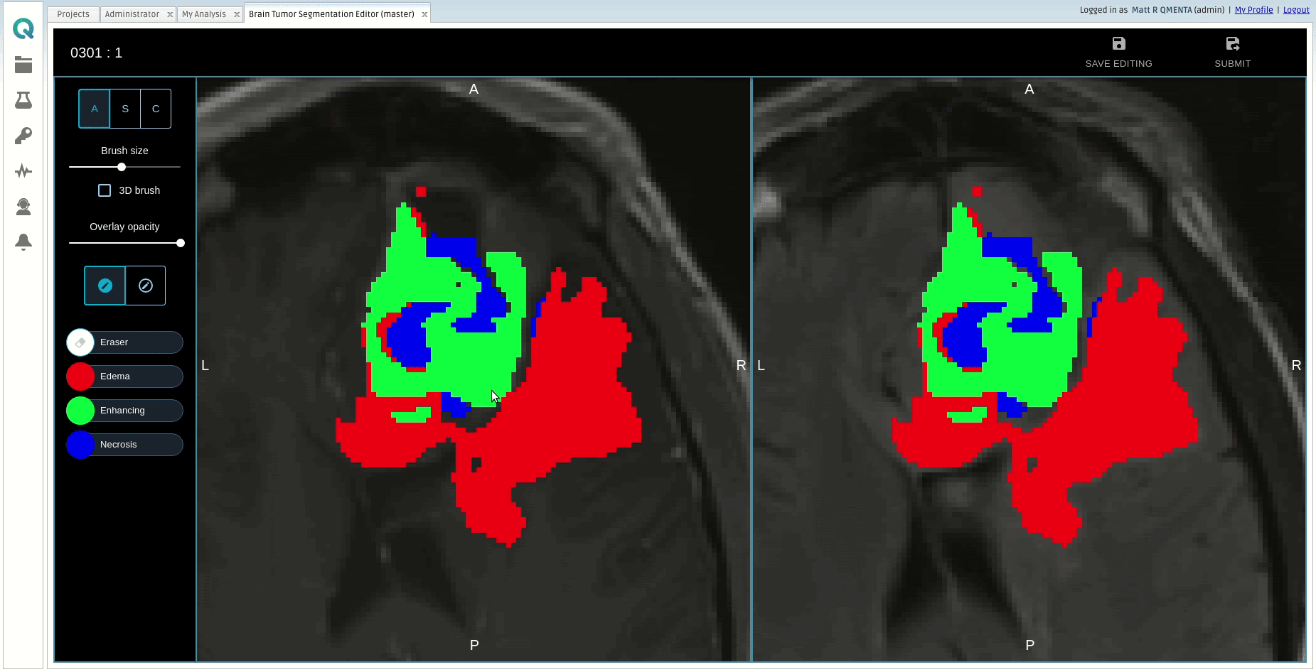
The segmentation map consists of the following value to region of interest mapping:The workflow expects a post-contrast T1 with Gadolinium agent (T1-Gad), a T2 FLAIR and optionally a native T1, and then produces class-confidence maps (softmaps) for each glioma sub-region, a segmentation map with the final prediction, and a PDF report providing a visual overview of the segmentation, tumoral volume quantification and a selection of radiomic features.
- 0: non-tumoral tissue and background
- 1: t2-lesion
- 2: enhancing
- 3: necrosis
The brain tumor segmentation tool included in the workflow is primarily made of a Convolutional Neural Network trained on a large proprietary database of pre- and post-operative MRI scans of patients with gliomas of varying shape, extension, and composition.
It includes an image pre-processing pipeline that resamples, co-aligns and skull-strips the input images to be prepared for inference with the aforementioned model. The model generates confidence-score maps for each tumoral sub-region and the final prediction map is obtained by assigning to each voxel the class with the highest confidence. Post-processing is applied after inference by default.
The output segmentation maps along with the preprocessed anatomical images are passed to a manual step in which the user can add, remove and correct the labels for each of the regions of interest.
Afterwards, the manually corrected segmentation map is used to compute volumes and ratios of volumes of the different regions of interest. The segmentation map is also used, together with the post-contrast T1 image, to compute the following radiomic features:
- First Order statistics
- Shape features
- Gray Level Cooccurence Matrix (GLCM)
- First Order statistics over Laplacian of Gaussian (LoG) with different sigma values (1mm, 3mm and 5mm).
All this volumetric and radiomic information is included in the PDF report. Volumetric information is added to the subject's session metadata.
If the inputs are DICOM, the outputs are converted to DICOM as well (see Output container files).
Required inputs
- T1 Gad: anatomical T1 post-contrast. Must be labeled as 'T1' modality and 'gad' tag.
- FLAIR: T2 FLAIR image. Must be labeled as 'T2' modality and 'flair' tag.
Optional inputs
- T1: T1 pre-contrast. Including a T1 pre-contrast image helps in the segmentation of necrotic tissue. Must be labeled as 'T1' modality and have no tags.
Minimum input requirements
- Whole-brain FOVs are recommended for all input modalities.
- 3D images are highly recommended. The preferred resolution is 1mm isotropic in all cases.
- If resampling and co-registration pre-processing steps are skipped, the input images must have the same resolution and voxel dimensions.
Settings
-
Image pre-processing
-
Isotropic resampling to 1mm (checkbox). Controls whether isotropic resampling to 1mm is executed or not. As the model was trained on images with such resolution, it is compulsory to have the inputs passed to the model at such resolution. Therefore, this option should be unchecked only when the input image has already 1mm isotropic resolution.
-
Co-registration of image modalities (checkbox). Controls whether the co-registration of input images is run or not. This step should be skipped only when the input images are already co-aligned, in order to save some processing time.
-
Skull stripping (checkbox). Controls whether the skull-stripping step should be run or not. The segmentation network was trained exclusively with skull-stripped data, so the inclusion of input images with skull would introduce confounding information that might worsen the results provided by the tool.
-
-
Image post-processing
- Keep connected components bigger than X size (checkbox). Controls whether the inital prediction of the model is filtered based on the size of the connected components.
- Specify minimum size X (voxel count) (integer). Specifies the minimum size in voxels that a component from the initial prediction should have in order to be kept. Goes along with the previous setting.
- Keep N biggest connected components (checkbox). Controls whether the inital prediction of the model is filtered by keeping only a certain number of (the largest) connected components.
- Specify number N of biggest connected components to keep (integer). Specifies the number of connected components to keep.Goes along with the previous setting.
Output container files
-
DICOM files: (Only when inputs are DICOM)
- T1 GAD T1_gad_aligned.zip: Pre-processed T1 Gadolinium contrast image
- PREDICTIONS RT STRUCT OVER T1 GAD predictions_RTSS_t1_gad.zip: Predictions of BTS as a DICOM RT Structure Set linked to the T1 GAD image
- T2 FLAIR T2_flair_aligned.zip: Pre-processed T2-FLAIR image
- PREDICTIONS RT STRUCT OVER T2-FLAIR predictions_RTSS_t2_flair.zip: Predictions of BTS as a DICOM RT Structure Set linked to the T2-FLAIR image
- T1 T1_aligned.zip (optional): Pre-processed T1 image
- PREDICTIONS RT STRUCT OVER T1 predictions_RTSS_t1.zip (optional): Predictions of BTS as a DICOM RT Structure Set linked to the T1 image
- PREDICTIONS (RGB) predictions_RGB.zip: Predictions of BTS as an RGB DICOM. It contains the predicted masks overlaid to the T1 GAD image.
- PREDICTIONS (SEG) predictions_SEG.zip: Predictions of BTS as a DICOM Seg object.
- REPORT REPORT.zip: pdf report from BTS Report box as an RGB DICOM. It can be displayed in a clinical viewer.
-
report.pdf: PDF report.
-
NIfTI files:
- MASK BRAIN brain_mask.nii.gz: Extracted brain mask, used for skull-stripping.
- SOFTMAP t2_lesion.nii.gz: Pseudo-probability map (softmap) of the lesion visible in the T2-FLAIR image.
- SOFTMAP enhancing.nii.gz: Pseudo-probability map (softmap) of the enhancing region.
- SOFTMAP necrosis.nii.gz: Pseudo-probability map (softmap) of the necrosis.
- LABELS prediction.nii.gz: Final segmentation map, with edema (1), enhancing tumor (2) and necrosis (3).
- T1 GAD t1_gad.nii.gz: Pre-processed T1 Gadolinium contrast used to perform inference on the DL model.
- T2 FLAIR t2_flair.nii.gz: Pre-processed T2-FLAIR used to perform inference on the DL model.
- T1 t1.nii.gz (optional): Pre-processed T1 used to perform inference on the DL model.
- preprocessing/
- t1_gad_aligned.nii.gz: Pre-processed T1 Gadolinium contrast before skull-stripping. Used for visualization of results.
- t2_flair_aligned.nii.gz: Pre-processed T2-FLAIR contrast before skull-stripping. Used for visualization of results.
- t1_aligned.nii.gz (optional): Pre-processed T1 before skull-stripping. Used for visualization of results.
- radiomics/
- LOG LoG_sigma_1mm_t1_gad.nii.gz: Laplacian of Gaussian of T1-Gad image with sigma=1mm.
- LOG LoG_sigma_3mm_t1_gad.nii.gz: Laplacian of Gaussian of T1-Gad image with sigma=3mm.
- LOG LoG_sigma_5mm_t1_gad.nii.gz: Laplacian of Gaussian of T1-Gad image with sigma=5mm.
- report/
- online_summary_report.html: Online report, visible in the fourth tab of "Show results".
- online_radiomics_report.html: Online report, visible in the fourth tab of "Show results".
- tumor_segmentation_over_reference_axial.png: Axial view of multiplanar representation of the segmentation map.
- tumor_segmentation_over_reference_coronal.png: Coronal view of multiplanar representation of the segmentation map.
- tumor_segmentation_over_reference_sagittal.png: Sagittal view of multiplanar representation of the segmentation map.
- tables/
- VOLUMES tumor_volumes.csv: CSV with the volume quantification of the different tumoral regions of interest.
- RATIOS tumor_volumes_ratios.csv: CSV with the volume ratios of the different tumoral regions of interest.
- RADIOMICS radiomic_features.csv: CSV with the extracted radiomic features.
References
- ANTs registration: Avants et al. 2008
- ROBEX skull-stripping: Iglesias et al. 2011
- CNN architecture: Puch et al. 2019
- Radiomic features: van Griethuysen et al. 2017
Create free account now!
