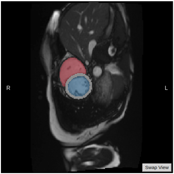
This tool uses a fully convolutional neural network to automatically segment the left ventricle, the left ventricle myocardium and the right ventricle of cine-MRI cardiac images from three possible views (short axis, four-chamber long axis and two-chamber long axis). It is based on the Github repository DLTK models: UKBB cardiac segmentation cine-MRI.

The tool detects the end-systolic and end-diastolic frames in the image and uses the information to compute a set of clinical values:
- Left ventricular end-diastolic volume (LVEDV)
- Left ventricular end-systolic volume (LVESV)
- Left ventricular myocardial mass (LVM)
- Right ventricular end-diastolic volume (RVEDV)
- Right ventricular end-systolic volume (RVESV)
The full workflow includes: - DICOM files to NIfTI conversion and reorientation to standard. - Cardiac segmentation using fully convolutional neural network.
Required inputs
- One or more cine-MRI cardiac images.
- The inputs must be labeled as either:
- cardiac short_axis 4d
- cardiac long_axis 2ch 4d
- cardiac long_axis 4ch 4d
Outputs
For each of the input images the following files are outputted:
- Original image in 4D
- Segmentation of the original image
- End-diastolic frame of the original image
- Segmentation of the end-diastolic frame
- End-systolic frame of the original image
- Segmentation of the end-systolic frame
- Clinical measures in a csv file
- Report for quality control purposes
Advanced settings
- DCM2NII:
- Preferred DICOM to NIfTI conversion tool (drop-down selection):
- DCM2niix (Default)
- Mrtrix
- DCM2nii
- diffunpack
- MRIConvert
- Preferred DICOM to NIfTI conversion tool (drop-down selection):
References
-
[1] W. Bai, et al. Human-level CMR image analysis with deep fully convolutional networks. https://doi.org/10.48550/arXiv.1710.09289
-
[2] S. Petersen, et al. Reference ranges for cardiac structure and function using cardiovascular magnetic resonance (CMR) in Caucasians from the UK Biobank population cohort. Journal of Cardiovascular Magnetic Resonance, 19:18, 2017. https://doi.org/10.1186/s12968-017-0327-9
Create free account now!
